
Fig. 1.
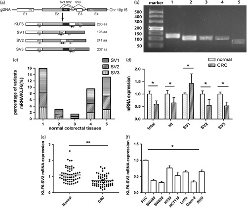
mRNA expression of KLF6-SV2 in CRC. Expression level was detected by qRT-PCR and calculated using the 2-ΔΔCt formula. (a) Gene structure of KLF6 and its splices variants. E1–E4 means exon 1–4. The arrows indicate the position of the primers used for PCR (Hanoun et al., 2010). (b) KLF6 and its splicing variants in normal colorectal tissue. PCR products were visualized after agarose gel electrophoresis. 1–5 stands for total KLF6, wild type KLF6, SV1, SV2, SV3. (c) Percentage of different variants mRNA expression in five normal colorectal tissues. (d) Different expression of KLF6 and alternative splicing in five pairs of normal and CRC tissues. Wt stands for wild type KLF6. (e) Different expression between CRC tumor tissue and adjacent tissue. (f) Different expression between FHC cell and seven CRC cell lines. **P<0.01, *P<0.05. CRC, colorectal cancer; KLF6, Kruppel like factor 6.