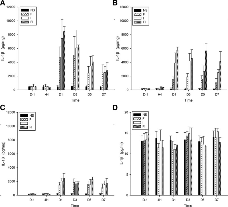
Figure 3.

The expression of interleukin-1β (IL-1β) in the spinal cord, bilateral dorsal root ganglion (DRG), and cerebrospinal fluid (CSF) after treatment of fentanyl and/or surgical incision. Sprague Dawley rats received subcutaneous fentanyl (60 μg/kg × 4) or normal saline (NS) and/or plantar incision (group NS = the control group; group F = rats received fentanyl only; group I = rats received NS and plantar incision; group FI = rats received fentanyl and plantar incision). Lumbar spinal cord (A), ipsilateral DRG (B), contralateral DRG (C) to surgical sites and CSF (D) of 4 rats were collected in each group. The expression of IL-1β (pg/mg for tissue, pg/mL for CSF) on the day before (D−1), at 4 h (H4) and on the days 1, 3, 5, 7 after drug injections (D1, D3, D5, and D7) were detected by enzyme-linked immunosorbent assay. All data are shown as mean ± standard deviation (n = 4).
