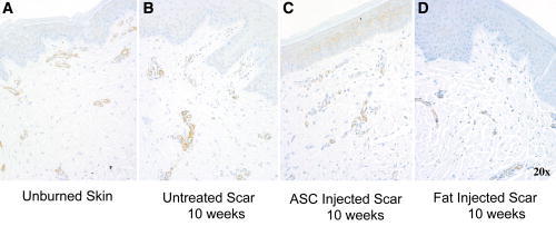
Fig. 10.

α-SMA antibody immunohistochemistry was performed with the myofibroblast staining brown. Unburned skin shows regularity of vessels at epidermal and dermal interface (A). Vertical vessels are again noted in the scars and amount of vascularity appears to be increased in untreated burns (B). Samples treated with autologous fat injections or ASCs at 10 weeks postinjection and 20 weeks postinjury appear to have decreased vessel penetration and presence (C, D).
