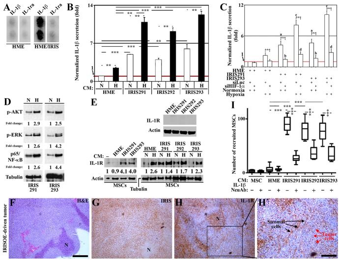
Figure 1. IRISOE TNBC cells secrete IL-1 β to recruit and activate MSCs.
(A) IL-1β and IL-1ra levels in HME cells transfected with doxycycline-inducible IRIS allele in the absence (HME) or presence of 2μg/ml of Dox (72h, HME/IRIS). (B) Normalized IL-1β level detected using ELISA in the conditioned medium (CM) of HME, IRIS291, IRIS292, or IRIS293 cells grown under normoxic (N) or hypoxic (H) conditions for 24h. (C) Normalized IL-1β level detected using ELISA in the CM of HME, IRIS291, or IRIS293 cells transfected with siLuc or siHIF-1α for 48h, followed by growth in N or H conditions for an additional 24h. (D) Western blot analysis for the level of activated AKT, ERK, or NF-κB/p65 in IRIS291, or IRIS293 cells grown under N or H conditions for 24h. (E) Western blot analysis of the surface expression of IL-R in naïve HME, IRIS291-IRIS293 (upper), naïve MSCs grown for 24h in the absence [-] or presence of CM from naïve HME, IRIS291, IRIS292, or IRIS293 pre-exposed or not to hypoxic conditions (lower). (F-H) IHC analysis of the expression of IRIS, and IL-1R in a 1° orthotopic IRISOE mammary tumor. (H) Higher magnification image of the area squared in I. Scale bar: 500μm in F-H, and 100μm in H`. (I) Recruitment of MSC towards IRISOE cells CM in the absence or presence of an IL-1β NeuAb analyzed using Boyden chamber.

