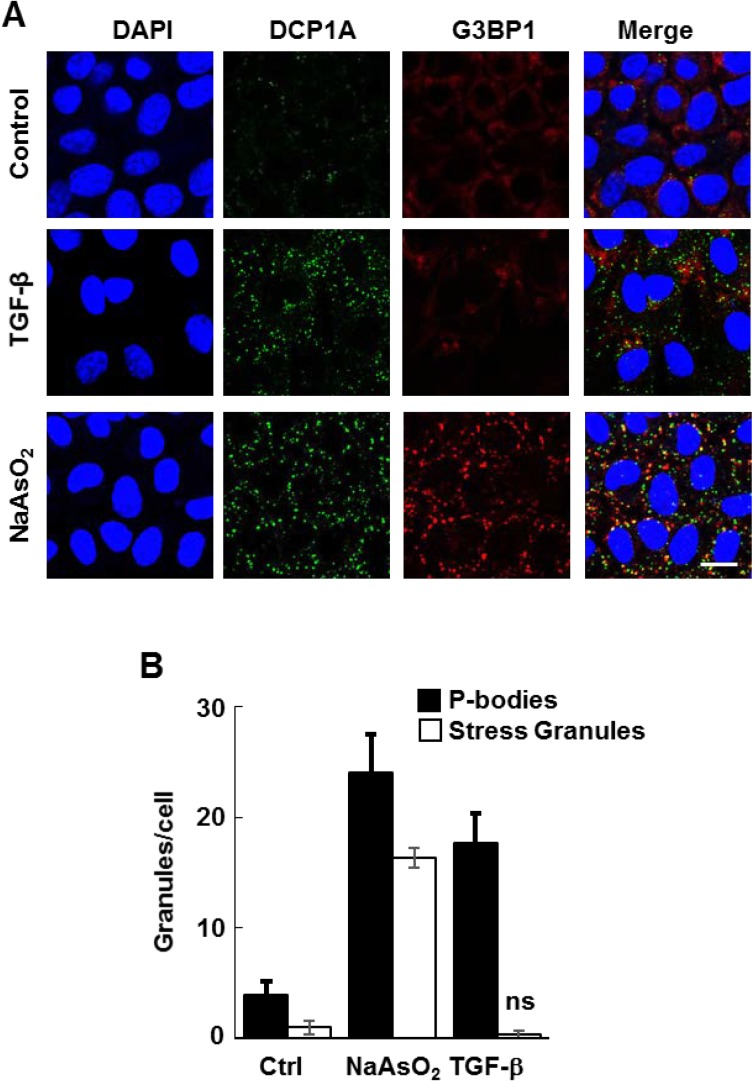
Figure 1. TGF-β induces the formation of P-bodies.
(A) NMuMG cells were treated without (Control) or with TGF-β for 24 h or sodium arsenite for 1 h, fixed and stained with anti-DCP1A (green) or anti-G3BP (red) to detect P-bodies or stress granules, respectively. Nuclei were stained with DAPI (blue). Bar, 10 μm. (B) The number of granules (P-bodies or stress granules) per cell was quantified (n > 100 cells per treatment). Data represents means ± SEM for triplicate experiments.

