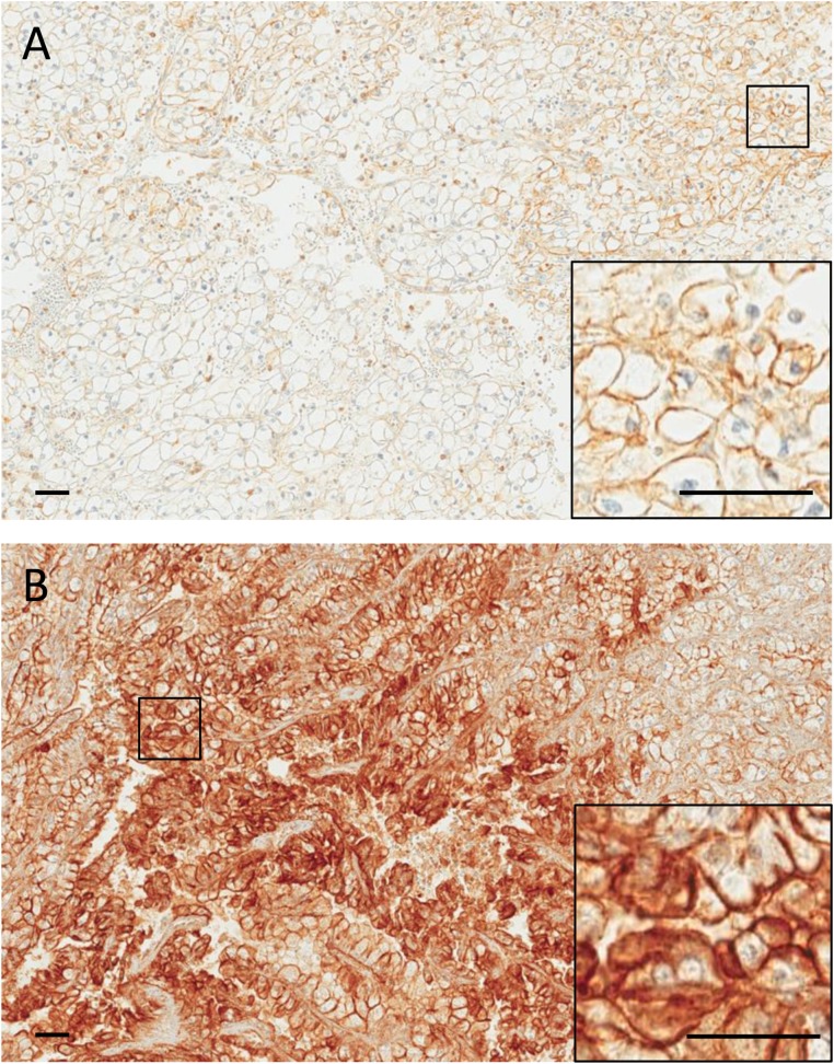
Figure 1.
Representative images of a primary ccRCC (A) and its corresponding metastasis (B) immunostained for c-Met. A, weak (1+) membranous staining is observed in a subset of tumor cells. B, intense (3+) membranous staining is observed in a large fraction of tumor cells. Insets show higher magnifications of the selected areas. Scale bars: 50 µm.

