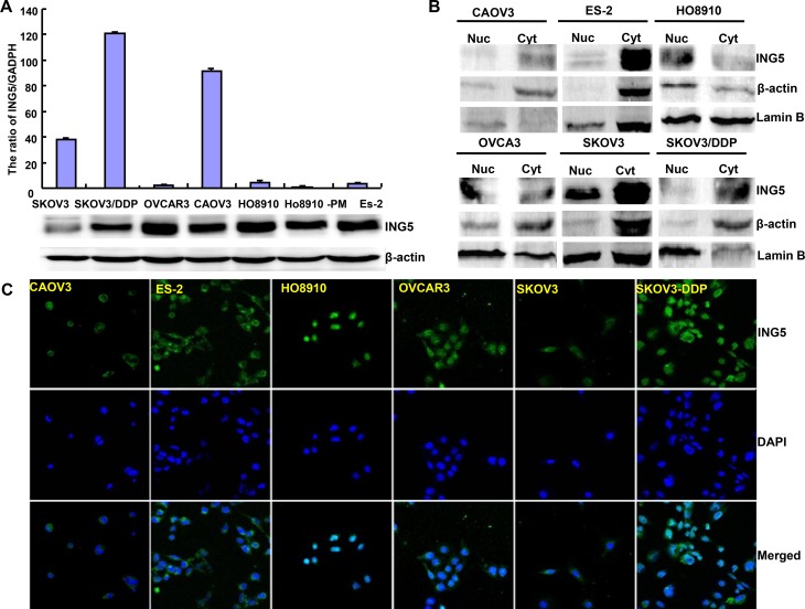
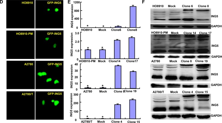
Figure 1. The expression and subcellular localization of ING5 in ovarian cancer cells.
Endogenous ING5 expression was screened in several kinds of ovarian cancer cells, including SKOV3, SKOV3/DDP, OVCAR3, OVCA3, HO8910, HO8910-pm, and ES-2, evidenced by RT-PCR (A), Western blot (A, B), immunofluorescence and nucleocytosolic fraction (C). After transfection of pEGFP-N1-ING5, ING5 expression became strong in HO8910, HO8910-PM, A2780 and A2780/T cells by immunofluorescence (D), RT-PCR (E), and Western blot (F). *p < 0.05, compared with transfectants.


