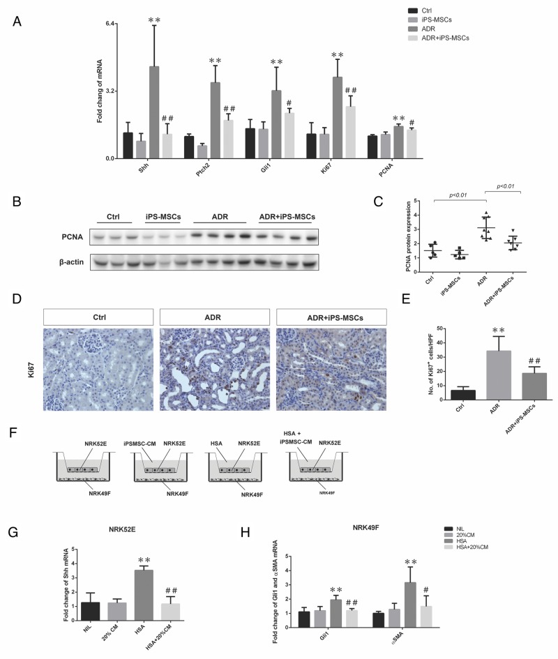
Figure 5. iPS-MSCs modulated fibroblast activation by attenuating hedgehog signaling pathway.
(A) Real-time PCR of Shh, Ptch2, Gli1, Ki67, and PCNA expression in the kidneys at 28 days of ADR injection (Ctrl: n=5; iPS-MSCs: n=5; ADR: n=8; and ADR+iPS-MSCs: n=7). (B), (C) Western blot to detect PCNA expression in renal cortex and its quantification. (D), (E) Immunohistochemical staining and quantification of Ki67 expression in tubulointerstitium. n=5 for Ctrl, n=8 for ADR and n=7 for ADR+iPS-MSCs. (F) Co-culture design to test the interaction of NRK52E and NRK49F and the role of iPS-MSC conditioned medium. (G). Real-time PCR of Shh mRNA expression in NRK52E (n=3). (H). Real-time PCR of Gli1 and αSMA mRNA in NRK49F collected from co-culture. Data were collected from three independent experiments. **p<0.01 versus Ctrl; #p<0.05 and ##p<0.01 versus ADR.

