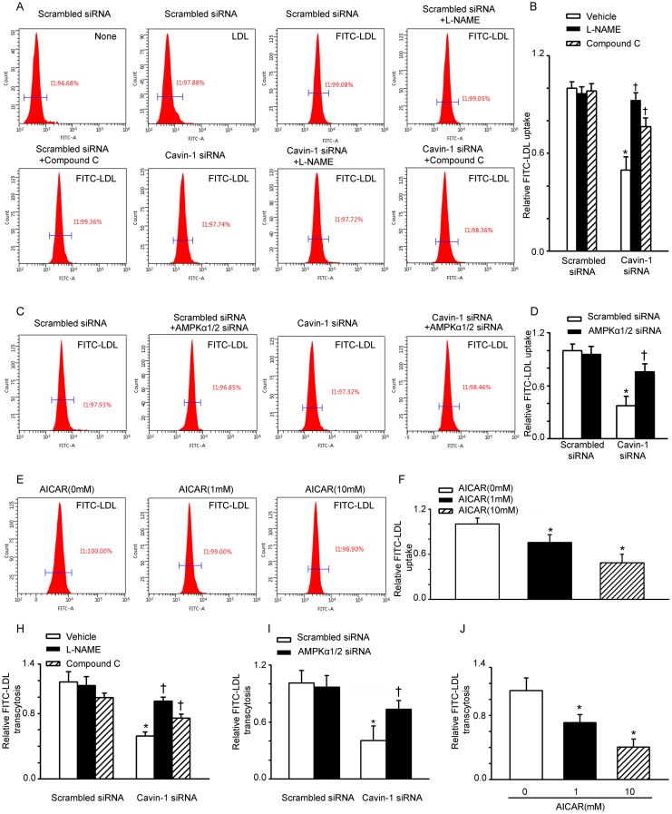
Figure 1. Cavin-1 siRNA suppressed LDL uptake and LDL transcytosis in HUVECs.
(A, B) Cells were transfected with scrambled siRNA or cavin-1 siRNA (20 nM) for 48 h, then incubated with L-NAME (50 μM) or Compound C (1 μM ) for 48 h. (C, D) Cells were transfected with scrambled siRNA (20 nM) or cavin-1 siRNA (20 nM) for 3 h, then co-transfected with scrambled siRNA (10 nM) or AMPKα1/2 siRNA (10 nM) for 45 h. (E-F) Cells were treated with indicated concentration of AICAR. (A, C, E) Flow cytometry images of FITC-LDL uptake in HUVECs incubated with FITC-LDL. (B, D, F) Summary bar graph showing the mean FITC-LDL fluorescent intensity in each group. (H, I, J) Quantitative summary of FITC-LDL transcytosis in HUVECs. * P <0.05 vs. scrambled siRNA or AICAR (0 mM); † P <0.05 vs. cavin-1 siRNA, n=4.

