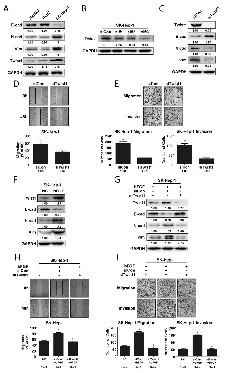
Figure 3. Twist1 played a crucial role in bFGF-induced EMT of HCC cells.
(A) Highly metastatic cell line SK-Hep-1 exhibited a higher extent of EMT and expressed more Twist1. (B) Knockdown efficiency of Twist1 siRNA in SK-Hep-1 cells was validated by western blotting. (C) E-cad, N-cad and Vimentin levels were detected by western blotting after silencing Twist1 in SK-Hep-1 cells. The metastatic potential of SK-Hep-1 cells after Twist1 knockdown was evaluated by wound healing (D) and migration and invasion assay (E). (F) bFGF upregulated Twist1 expression in SK-Hep-1 cells. (G) Western blotting was used to validate cell phenotypes associated with EMT in Twist1-knockdown SK-Hep-1 cells treated with bFGF. The metastatic potential of Twist1-knockdown SK-Hep-1 cells treated with bFGF was evaluated by wound healing (H) and migration and invasion assay (I), and Twist1 was found to be important for EMT induced by bFGF. *p<0.05 compared with control, #p<0.05 compared with bFGF.

