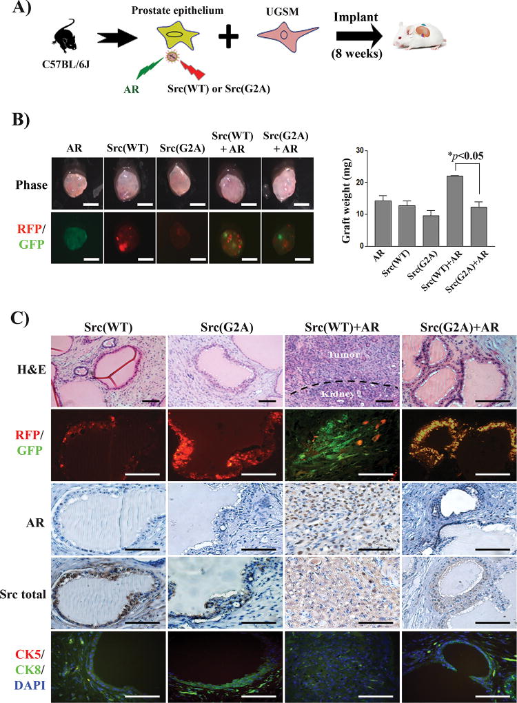
Figure 4. Loss of myristoylation in Src kinase inhibits the synergy of Src(WT) with androgen receptor (AR) in prostate tumorigenesis.
(A) Schematic for examining the synergy of Src and AR in prostate tumorigenesis. Primary prostate epithelial cells were transduced with AR (GFP marker), Src(WT) (RFP marker), Src(G2A) (RFP marker), or co-transduced with Src(WT)/Src(G2A) and AR and the infected cells were combined with UGSM, and implanted under the renal capsule of SCID mice. Regenerated prostate tissue was isolated after 8 weeks. (B) Representative images of regenerated prostate tissue and RFP/GFP detection (scale bar, 2 mm). The weight of prostate tissues was compared in the bar graph. The * indicates an unpaired, two-tailed t test. (C) H&E, RFP/GFP, and IHC staining of AR and total Src, and co-staining of CK8, CK5 and DAPI in regenerated tissue. Scale bar, 100 µm.

