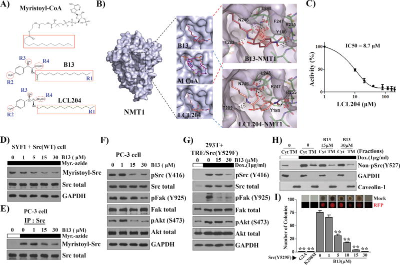
Figure 6. Myristoyl-CoA analog, B13, inhibits NMT1 enzymatic activity and suppresses Src kinase mediated cell transformation.
(A) Comparison of the myristoyl-CoA structure with B13 and LCL204. The C14 acyl-group (R1) highlighted with a red square in B13 is identical with that in myristoyl-CoA. R2 (nitro group), R3 (hydroxyl group), and R4 (hydroxymethyl group) are important for the inhibitory effect based on structure-activity relationship (SAR) analysis in the Supplemental Figure S9. (B) The analysis of the NMT1 protein structure with B13 and LCL204 inhibitor. The binding site of myristoyl-CoA with the NMT1 crystal structure was replaced with B13. The upper right panel shows B13 interactions with neighboring side chains. Myristoyl-CoA and LCL204 were modeled in the NMT1 binding pocket. The bottom right panel shows that LCL204 interactions with the neighboring side chains. According to the structure replacement, the replacement of the methoxide group in B13 with -CH2 led to a B13 derivative, LCL204, which favors the interactions of the functional groups with amino acids in the binding pocket of NMT1. (C) The inhibitory effect of LCL204 on NMT1 enzymatic activity. The IC50 of LCL204 on NMT1 enzymatic activity was 8.7 µM, which increased about 9-fold in comparison with that of B13. (D–E) SYF1 cells were transduced with Src(WT) and the transduced cells, SYF1+Src(WT) (D) or PC-3, (E) were cultured with B13 overnight followed by myristic acid-azide (60 µM) for 8 h. The lysates from SYF1+Src(WT) cells (D) and Src immunoprecipitates from PC-3 cells (E) were subjected to Click chemistry. The expression levels of myristoyl-Src detected by streptavidin-HRP, total Src, and GAPDH were analyzed by immunoblotting. (F) The expression levels of the indicated proteins in the total lysates from (E) were measured by immunoblotting. (G) 293T cells expressing doxycycline inducible Src(Y529F) were treated with/without doxycycline (Dox, 1µg/ml) and with 0 (DMSO), 15, and 30 µM of B13 for 24 h. The levels of the indicated proteins were determined by immunoblotting. (H) The lysate from (G) was fractionated. Caveolin-1 and GAPDH were used as markers for total cytoplasmic membrane (TM) and cytosolic fraction (Cyt), respectively. Immunoblot detection using the non-pSrc(Y527F) antibody represented the de novo synthesized Src kinase. (I) SYF1 cells were transduced with Src (Y529F), Src (Y529F/G2A), or Src (Y529F/K298M) and subjected to the soft agar assay. SYF1-Src(Y529F) cells were treated with B13 and the number of resulting colonies was counted. Representative phase and RFP images of colonies in the soft agar assay are displayed. **: P<0.01.

