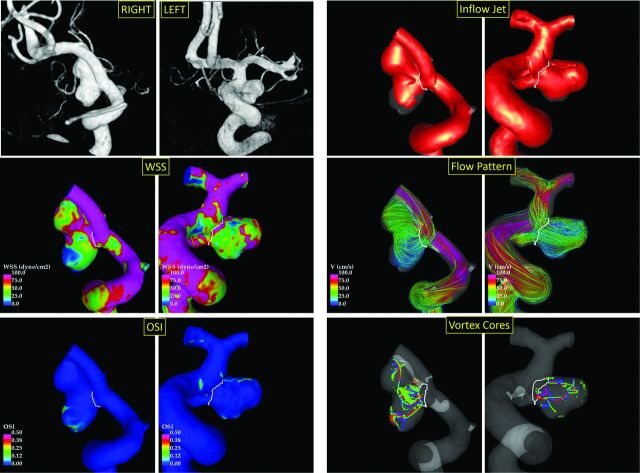
Fig 3.
Example of ruptured (right posterior communicating artery aneurysm, left column) and unruptured (right posterior communicating artery aneurysm, right column) mirror aneurysm pairs. The Left panel shows from top to bottom: 3D rotational angiography images, WSS distributions, and OSI distributions. The right panel shows from top to bottom: inflow jets, flow patterns, and vortex core lines at 4 times during the cardiac cycle.

