
Figure 4. Spt5V1008D ESCs show defects in differentiation.
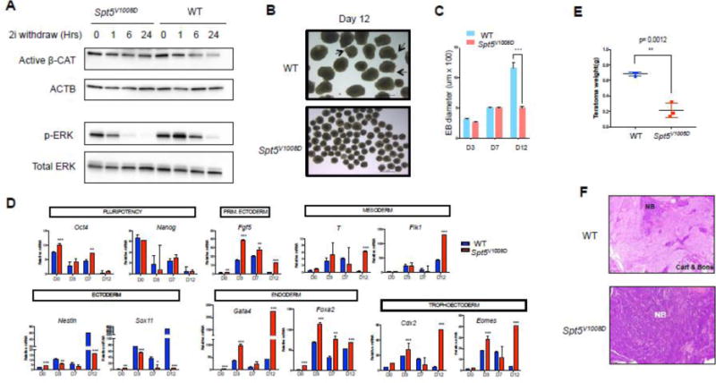
(A) Western blots comparing the activity of Wnt (active β-Catenin) and ERK signaling (p-ERK) between wild-type and mutant ESCs after removal of GSK3 and ERK inhibitors (2i) from the serum free media for different time length (Hrs: hours) as indicated. β-actin (ACTB) and total ERK serve as loading controls.
(B) Bright field imaging of day-12 embryoid bodies (EBs) from wild-type and mutant ESCs. Arrows depict cystic structures seen in wild-type day-12 EBs.
(C) Comparison of mean diameters (uM) of 20 EBs at indicated days of differentiation (n=3, mean ± SEM, ***p<0.001).
(D) Quantitative RT-PCR comparing mRNA levels of pluripotency and differentiation marker genes between wild-type and mutant EBs. Gene expression is normalized to Gapdh and presented as fold change relative to day0 (n=3, mean ± SEM, * p<0.05, **p<0.01, ***p<0.001).
(E) Teratoma weight comparison at time of collection. Data represents the average from three tumors in each group.
(F) Representative histology images of H&E stained teratoma sections. NB: undifferentiated neuroblastic tissue. Cart: cartilage. Note the teratoma derived from mutant ESCs (bottom) is full of NB with no signs of mature tissues (bone and cartilage).