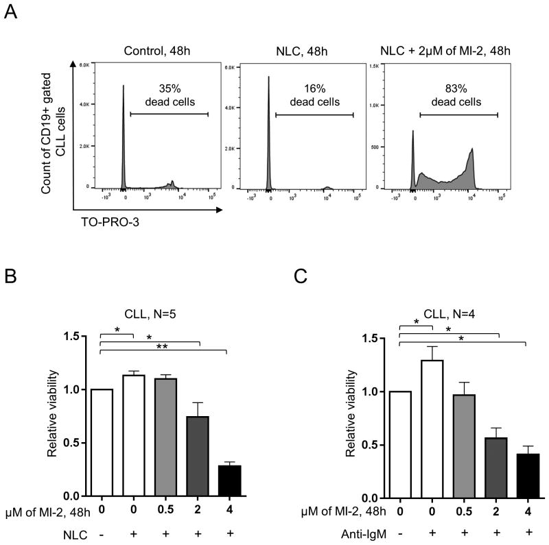
Figure 3. MI-2 overcomes the protective effect of the microenvironment.
(A) A representative flow panel showing the increase in cell death (TO-PRO-3 stained) in CD19 gated CLL cells, following exposure to 2 μM of MI-2 for 48h in the presence or absence of NLC. (B) PBMCs of 5 patients with CLL were incubated for 48h with 0, 0.5, 2, and 4 μM of MI-2 in the presence or absence of NLC. Shown is the viability of CD19 gated CLL cells as in (A). (C) Purified CLL cells (CD19 selection) (N=4) were incubated for 48h with 0, 0.5, 2, and 4 μM of MI-2 in the presence or absence of 5 μg/ml of anti-IgM. Shown is the CLL cells viability relative to untreated control without anti-IgM, measured by MTS. Error bars represent SEM.
SEM, standard error of mean; NLC, nurse-like cells; -, absence; +, presence; *, P<0.05; **, P<0.01.

