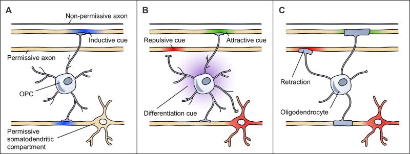
Figure 1. Different mechanisms of axon selection could produce the same CNS myelination patterns.
These three models are non-exhaustive and not mutually-exclusive. Structures with suprathreshold diameters are shown in yellow as permissive. The first axon has a subthreshold diameter, rendering it non-permissive. (a) An inductive cue (blue) turns on in axons to initiate their myelination. Shown here, an OPC senses a threshold myelination need from the second and fourth axons that causes the OPC to differentiate and myelinate these axons. These axons are among the permissive structures. (b) OPCs respond to attractive (green), permissive (yellow), and repulsive (red) cues on axons and other cellular compartments that regulate the contacts that OPCs make and the eventual placement of myelin sheaths. The OPC processes retract from repulsive cues on the third axon and the somatodendritic compartment. Upon differentiation, this cell will myelinate the second and fourth axons. The differentiation cue (purple) is uncoupled from axon selection. (c) OLs respond to attractive (green), permissive (yellow), and repulsive (red) cues that affect sheath initiation or stability. Retraction of an early sheath in response to a repulsive cue on the third axon is shown. This OL myelinates the second and fourth axons.

