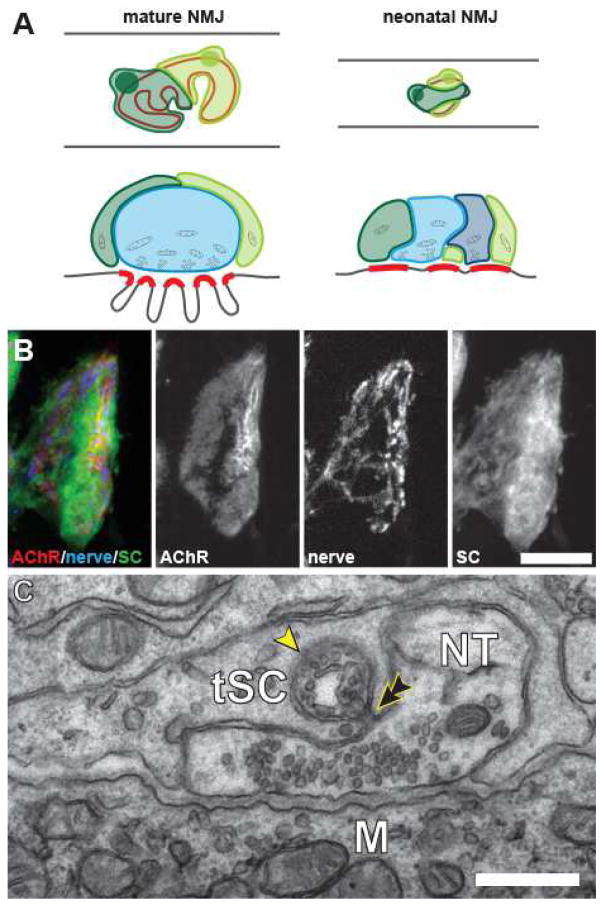
Figure 1. The arrangement of the cellular components at mouse neuromuscular junction.
(A) At mature NMJs, each tSCs (shown in shades of green) covers a non-overlapping portion of the nerve terminal (blue) that in turn covers the postsynaptic AChRs (indicated in red). At neonatal NMJs, this partitioning of terminals by tSCs has not yet been established and these cells interdigitate. Processes of the neonatal tSCs (in shades of green) make synapse-like apposition to the portions of the postsynaptic AChR aggregates unoccupied by the immature nerve terminals (in shades of blue). (B) Confocal images of the postsynaptic AChR aggregate (AChR), the motor axon terminals (nerve) and Schwann cells (SC) at a neonatal NMJ. As illustrated in (A), a significant portion of the oval-shaped AChR aggregate is not apposed by the innervating motor axon terminals, but rather by the processes of tSCs. (B) Electron micrograph of a single section in a serial series showing a tSCs consuming a portion of the presynaptic motor axon terminal (NT) that is still in contact with the target muscle fiber (M). A phagocytic vesicle containing components of the nerve terminal (arrowhead) is situated within tSC cytoplasm and has almost completely pinched off (double arrowhead) from this nerve terminal. Scale bars: 10 μm in (B) and 500 nm in (C).

