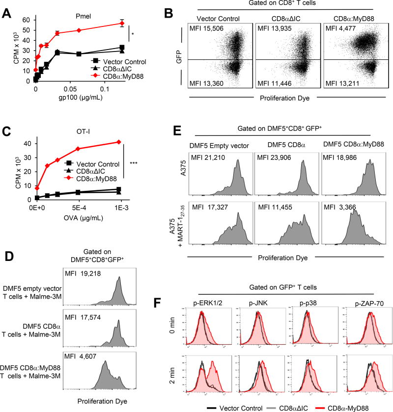
Figure 1. CD8α:MyD88 expression enhances T cell responses to tumor antigen.
A) Pmel T cells were engineered with the empty vector control, CD8αΔIC or CD8α:MyD88. T cell proliferation was measured by 3H-thymidine incorporation after stimulation with gp10025–33-pulsed splenocytes for 48 hours. B) Flow cytometry of pmel T cells co-cultured with splenocytes pulsed with 0.12 µg/mL gp10025–33 peptide at 72 hours. C) Proliferation of OT-I T cells co-cultured with OVA (SIINFEKL)-pulsed splenocytes measured by 3H-thymidine incorporation at 48 hours. D–E) DMF5 empty vector control, DMF5 CD8α or DMF5 CD8α:MyD88 human T cells were labeled with proliferation dye and cultured with HLA-A2+ MART-1+ Malme-3M or with HLA-A2+ MART-1− A375 melanoma pulsed or not with 10µg/ml/106 cells MART-127–35 cells at a 1:1 ratio. T cell proliferation was measured by flow cytometry after 5 days. F) Intracellular staining of phosphorylated proteins in transduced T cells stimulated with peptide-pulsed splenocytes and fixed at given timepoints. Values and error bars represent mean ± s.e.m. * p ≤ 0.05, ** p ≤ 0.01, *** p≤ 0.001 A and C, one-way ANOVA with Tukey’s Multiple Comparison Test; n=3 experimental replicates; representative of at least two independent experiments. D and E are representative of three independent experiments.

