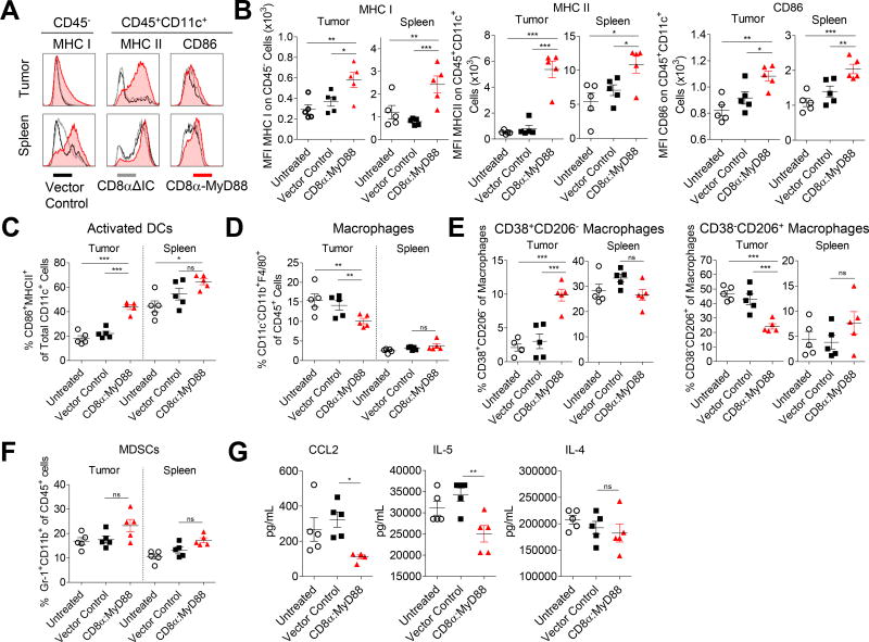
Figure 4. CD8α:MyD88 T cells alter the tumor microenvironment.
A–G) Mice bearing an established B16-F1 tumor (~50mm2) were exposed to a sublethal dose of irradiation (550 rads) followed by transfer of the CD8α:MyD88 or vector control pmel T cells (6×10⁶) by intraveneous injection one day later. Tissues were harvested and analyzed by flow cytometry one week after T cell transfer. Transduction efficiency of CD8α:MyD88 T cells was 50%. Each data point represented one mouse. A) Median fluorescence intensity (MFI) of MHC I on CD45− cells and MHC II and CD86 on CD11c+ dendritic cells (DCs). B and C) Frequency of CD11c+ DCs co-expressing activation markers CD86 and MHC class II in the tumor and spleen. D) Frequency of macrophages in the tumor and spleen as defined by CD11c−CD11b+F4/80+ cells. E) Frequency of M1 (CD38+CD206−) and M2 (CD38−CD206+) macrophages in the tumor and spleen. F) Frequency of myeloid-derived suppressor cells (MDSCs) in the tumor and spleen as defined by co-expression of Gr1 and CD11b. G) Luminex analysis of CCL2, IL-4, and IL-5 in tumor homogenate.

