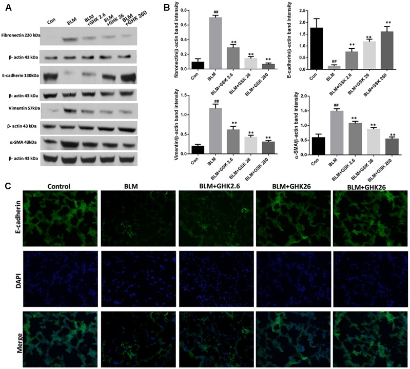
FIGURE 4.
Effects of GHK on BLM-mediated EMT in mice with BLM-induced fibrosis. (A) Fibronectin, E-cadherin, vimentin, and α-SMA protein expression, as measured by western blot analysis, in lung tissues in each group. (B) The densitometry values for the proteins were normalized to those of β-actin. (C) Immunofluorescence staining was performed to detect E-cadherin protein expression (green) in lung tissue sections in each group. The nuclei were stained by DAPI (blue). Representative images of each group are shown. All data represent the mean ± SEM of three independent experiments performed in triplicate. Statistical analysis was performed by one-way ANOVA and Turkey’s multiple-comparison test; compared with control group, ##P < 0.01; compared with BLM group, ∗P < 0.05, ∗∗P < 0.01.

