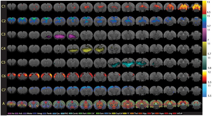
Figure 8.
Full spatial maps of resting-state networks in awake rats. Components (C1–C7) are ordered according to their reproducibility degree. Component 1 has significant cerebellar structures; Component 2 includes medial and lateral cortical structures resembling the human default mode network; Component 3 includes a basal-ganglia-hypothalamus network; Component 4 encompasses basal-ganglia-thalamus-hippocampus circuitry; Component 5 represents an autonomic pathway; Component 6 represents the sensory network; and Component 7 groups interoceptive structures to form a network. All components have been thresholded according to a mixture model approach. See Methods section for details. The atlas is based on the Paxinos and Watson Atlas (1998). Abbreviations: Ins, Insula; AcB, Nucleus Accumbens; Motor, Motor Cortex; Amyg, Amygdala; Parab, Parabrachial; CPu, Caudate-Putamen; PAG, Periaqueductal Gray; Cereb, Cerebellum; ParA, Parietal Association Cortex; Cnf, Cuneiform Nucleus; Som, Somatosensory Cortex; Ent, Entorhinal Cortex; Sup Coll, Superior Colliculus; FC, Frontal Cortex; Thal, Thalamus; TpA, Temporal Association Cortex; Hypo, Hypothalamus; cing, Cingulate Cortex (anterior and retrosplenial); Inf Coll, Inferior Colliculus. Figure reprinted with permission from Becerra et al. (2011b).

