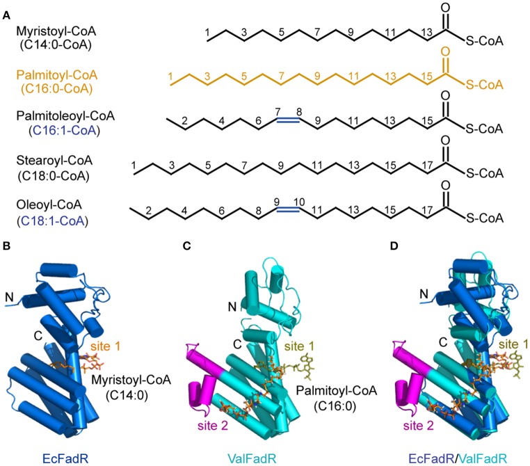
Figure 6.
Chemical structures of the representative FadR ligands and structural comparison of the liganded-ValFadR with the EcFadR-ligand complex structure. (A) Chemical structures of LC fatty acyl-CoAs, the ligands for FadR regulatory proteins. The palmitoyl-CoA ligand that was successfully crystalized into the FadR protein is highlighted in orange, whereas the double bond is indicated in blue. (B) Complex structure of the monomeric form of EcFadR liganded with myristoyl-CoA ligand. The architecture of the EcFadR (PDB: 1H9G) is shown in blue cylinder, whereas the ligand of myristoyl-CoA is given in orange stick. (C) Complex structure of the monomeric form of ValFadR liganded with palmitoyl-CoA ligand (PDB: 5DV5). The protomer form of ValFadR is shown in cyan cylinder, and the extra-40aa insert is given in magenta. The ligands are shown in sticks, one of which is of light green in the binding site 1 and the other one present in site 2 is indicated in red. (D) Superposition of the monomeric structure of ValFadR-ligand and EcFadR-ligand structure via their C-terminal ligand-binding domains.

