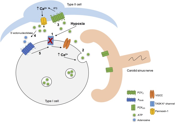
FIGURE 3.
Schematic representation of the CB “tripartite” synapse model proposed by Nurse and collaborators. Hypoxia induced type I cell depolarization through the inhibition of TASK1/3 K+ channels (1), leading to Ca2+ entry via voltage-gated Ca2+ channels (VGCC) and to ATP release (2). ATP excites postsynaptic P2X2/3 receptors on petrosal nerve terminal. ATP can also stimulate P2Y2 receptors in adjacent type II cells (3), leading to the Ca2+ release from intracellular stores via inositol triphosphate (IP3) signaling pathways and opening of pannexin-1 channels. This results in ATP release that could be break down by extracellular 5’ectonucleotidase into adenosine (4) (Conde et al., 2012a; Salman et al., 2017). Adenosine stimulates A2A adenosine receptors in type I cells, leading to the inhibition of TASK1/3 K+ channels, that enhance type I cell depolarization (5) (Xu et al., 2005) and, therefore ATP release. It is not represented but hypoxia stimulates adenosine release per se from type I cells (Conde and Monteiro, 2004) and high levels of ATP could inhibit pannexin-1 channels in type II cells and inhibit the chemoreceptor function via P2Y1 receptors, through a negative feedback mechanism. Adapted from Zhang et al. (2012), Nurse (2014), Murali and Nurse (2016).

