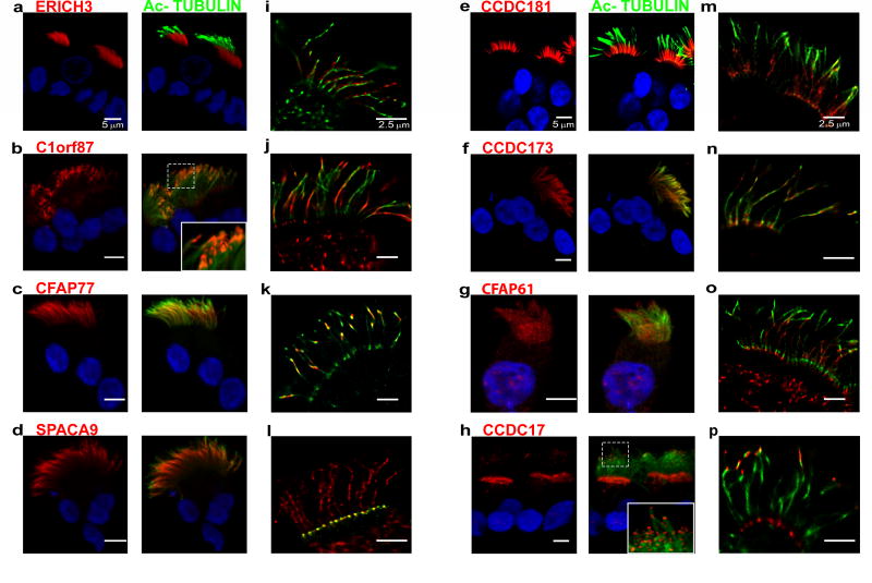
Figure 4.
Immunofluorescence localization of the indicated proteins in isolated ciliated cells (1st column; red) co-localized with Ac-TUB (2nd column; green) by conventional confocal microscopy. The 3rd column shows localization of the proteins in micrographs obtained using GSD microscopy. See text for details. Scale bar = 5 µm in left and center panels; nuclear staining was with Hoechst 33342 (blue). Scale bar = 2.5 µm in right panels.

