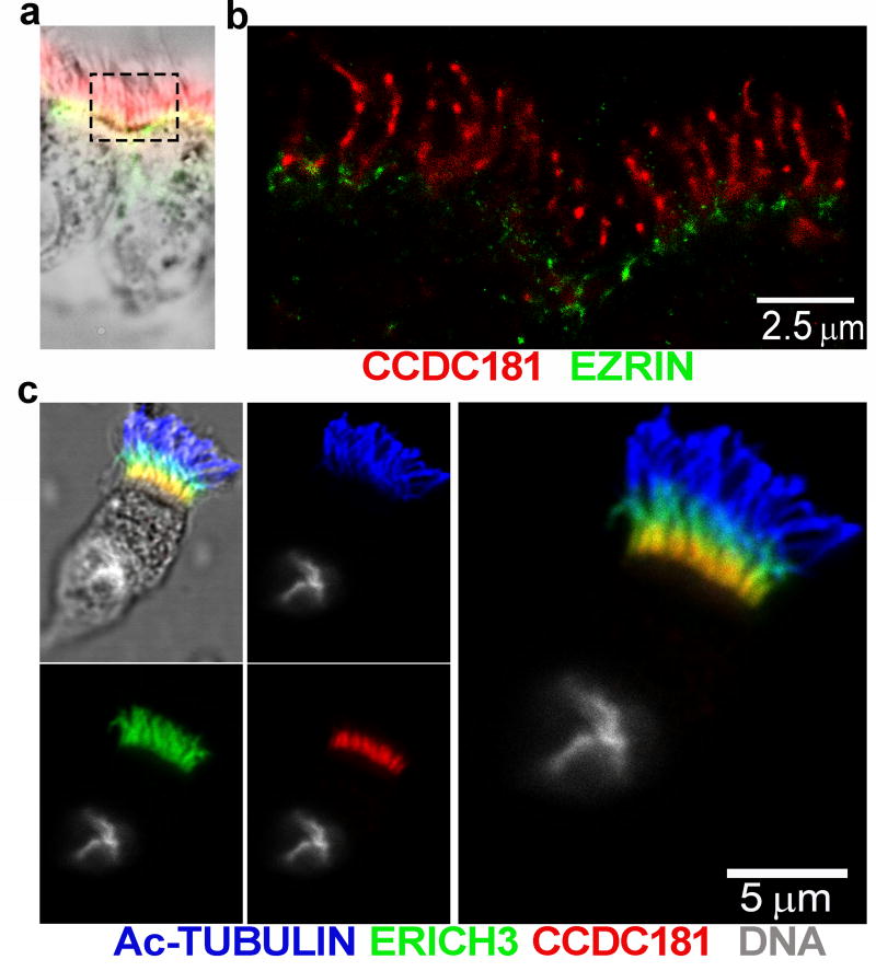
Figure 5.
a) Localization of CCDC181 (red) and ezrin (green) confirms that CCDC181 is a component of the ciliary axoneme and not the microvilli. A low magnification bright field image of the stained ciliated cells (a) imaged with super resolution microscopy (b) showing that CCDC181 localizes specifically to the cilia (scale bar = 2.5 µm). c) Co-localization of Ac-TUB (blue), ERICH3 (green), and CCDC181 (red) by immunofluorescence localizes CCDC181 to the proximal portion of airway cilia. Cell nuclei were stained with Hoechst 33342 (gray), scale bar = 5 µm.

