
Figure 5.
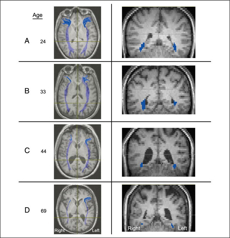
Representative axial (left) and coronal (right) slices taken from four participants, aged 24, 33, 44, and 69 years, revealing the loss of fibers as a function of increasing age, especially in the right hemisphere. Note that radiological convention is adopted with the right hemisphere depicted on the left of the image and vice versa.