
Figure 6. Pharmacological manipulations differentially regulate β1AR-mediated PKA activation on the Golgi.
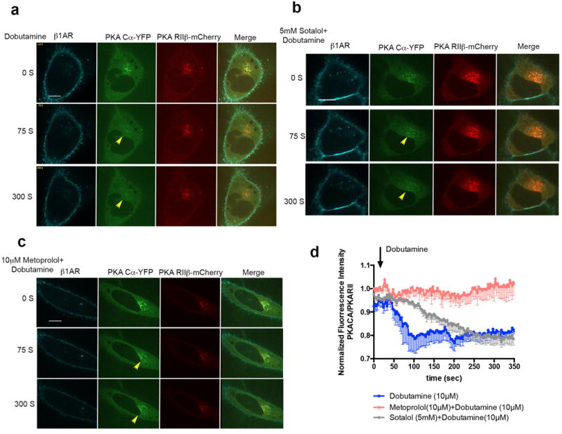
a. Confocal image frames of β1AR expressing HeLa cells treated with 10 μM dobutaine at indicated time points (n= 7 cells, 4 biological replicates). b. Confocal image frames of β1AR expressing HeLa cells pre-treated with 5mM sotalol and upon addition of 10μM dobutaine at indicated time points (n= 9 cells, 4 biological replicates). c. Confocal image frames of β1AR expressing HeLa cells pre-treated with 10μM metoprolol and upon addition of 10μM dobutamine at indicated time points (n= 5 cells, 4 biological replicates). d. Kinetics of PKA Cα-YFP translocation from the Golgi membrane in cells treated with 10μM dobutamine (blue), 10μM metoprolol+10μM dobutamine (red) or 5mM sotalol+ 10μM dobutamine; the fluorescent intensity of PKA Cα-YFP was normalized by PKA RIIβ-mCherry fluorescent (n= 7, 5 and 9, 4 biological replicates) (see methods).Scale bars, 10 μm.