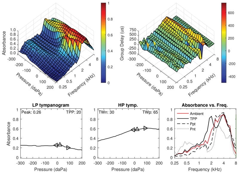
Figure 2.
Left ear of a 35 month-old female with DS, who was diagnosed with OME and had elevated air conduction hearing levels. DPOAEs were absent from 1–3 kHz, and present from 4–8 kHz. Standard 0.226-kHz tympanometry (not depicted) showed low-normal static admittance (0.2 mmho) and normal tympanometric width (81 daPa). Top left: Tympanometric absorbance (downswept pressure). Top right: Tympanometric GD. Bottom left: Average low-frequency tympanometry. Bottom middle: Average high-frequency tympanometry. Bottom right: Absorbance by frequency plots (at ambient, TPP, and the positive and negative tympanogram tails.)

