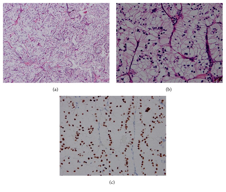
Figure 3.
Pathological findings. (a) The tumor cells were polygonal of a papillary carcinoma with clear cells and cells with granular eosinophilic cytoplasm (a) (HE staining, 100x magnification) and (b) (HE staining, 400x magnification). (c) The tumor cells in the kidney were visualized by immunohistochemistry staining for TFE3 which revealed positive staining of the nuclei.

