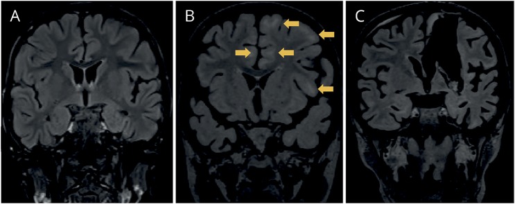
Figure 1. Coronal MRI images showing the evolution of white matter abnormality and atrophy of patient 1.
MRI (fluid-attenuated inversion recovery, FLAIR) in February 2013 (A), in September 2013 (B), and after left vertical parasagittal hemispherotomy in October 2013 (C). The arrows in B show subcortical regions with white matter FLAIR signal abnormality. Note also the progressive atrophy of the brain and the left temporal lobe in particular.

