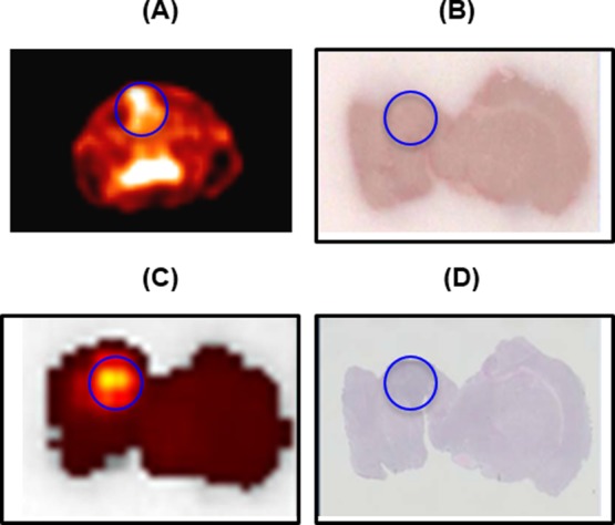
Figure 4.

(A) Transverse PET summed image (0–15 min) of the brain. (B) Post-mortem brain slice. (C) Optical image of the brain slice shown in B. (D) Immunohistochemical (H&E) staining of the brain slice B. The circle indicates the location of the tumor.
