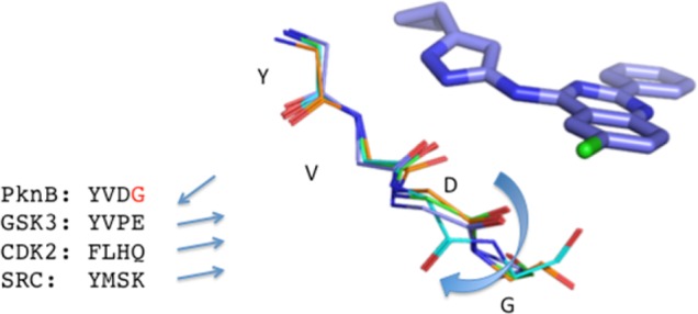
Figure 1.

Compound 22 docked into the ATP binding site of PknB, and the mammalian kinase GSK3β, CDK2, and SRC. Labels identify PknB residue numbers corresponding to cyan structure. For clarity, amino acid side chains are not shown. Straight arrows indicate the direction of the C=O bond between the 3rd and 4th residues in the sequences shown, and the curved arrow indicates flipping of the peptide bond between D and G residues. Alignment was done using C-alpha atoms. Red G is the residue that allows PKnB to undergo conformational change.
