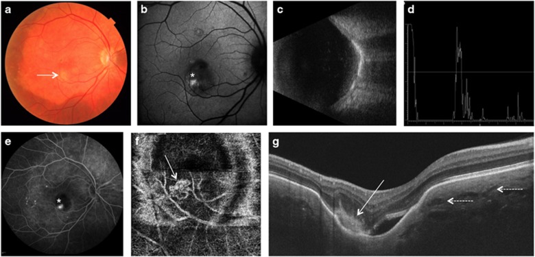
Figure 1.
Multimodal imaging of right eye in case 1. Color fundus photograph (a) showed a large choroidal osteoma at the posterior pole of the right eye. A more intense yellowish material was noted in the fovea (arrow) which was hyperautofluorescent (asterisk) on blue fundus autofluorescence (b). B scan echography (c) showed a solid highly reflective choroidal mass with acoustic shadowing. A scan ultrasonography (d) showed high intensity echo spike with sound attenuation posterior to the lesion. Axial length on A scan was 24.00 mm. Fluorescein angiography (e) showed an active leaking juxtafoveal choroidal neovascularization (CNV) which was visible on optical coherence tomography angiography (f), (arrow). Enhanced depth imaging optical coherence tomography (g) showed a choroidal sponge-like pattern with the presence of multiple intralesional layers (dashed arrows); in correspondence of the CNV an ill defined hyperreflective material was found on the slope of the choroidal excavation (arrow).

