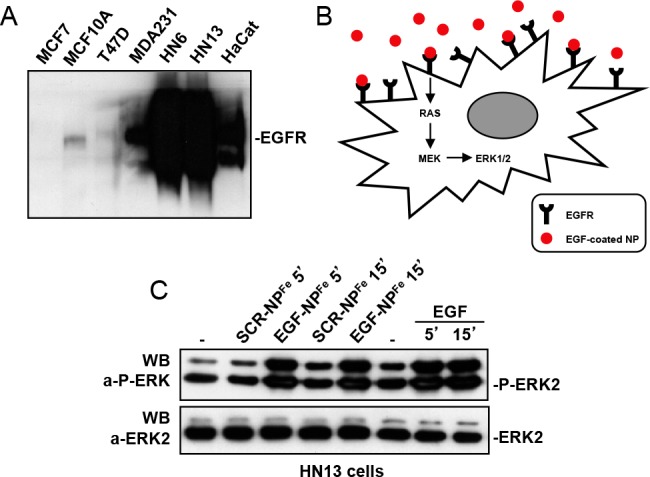
Figure 1.

(A) Western blot analysis, using an anti-EGFR antibody, of total cell lysates from breast cancer (MCF7, MCF10A, T47D, and MDA231) and head and neck squamous cell carcinoma (HN6, HN13) cells. HaCat are Human immortalized keratinocytes. (B) Schematic representation of the signaling pathway activated by EGF-NPFe upon interaction with the specific EGFR on target cells. (C) Measurement of ERK1/2 activation by Western blot analysis of NH13 total cell lysates, using a specific antiphospho-ERK1/2 antibody. An anti-ERK2 antibody was used in the lower panel for normalization purposes.
