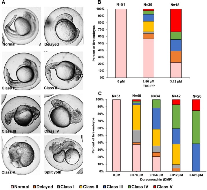
Figure 2. TDCIPP-exposed embryos phenocopy embryos exposed to dorsomorphin from 0.75 to 24 hpf.
(A) Representative 24-hpf images of normal embryos, delayed embryos, Class I–V levels of dorsalization, and split yolk embryos following exposure to TDCIPP or DMP. (B) Distribution of dorsalization classes following exposure to TDCIPP from 0.75 to 24 hpf. (C) Distribution of dorsalization classes following exposure to DMP from 0.75 to 24 hpf. Depending on the TDCIPP concentration (1.56 or 3.12 µM), the severity of dorsalization varied from mild (Class I) to strong (Class V).

