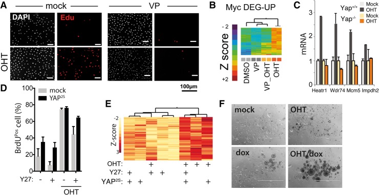
Figure 2.
Myc-driven cell cycle entry depends on YAP activity and cytoskeletal tension. (A–E) Serum-starved subconfluent fibroblasts were kept in low serum and treated as indicated. (A) Immunofluorescence analysis of Myc-induced cell cycle entry of 3T9MycER measured as EdU incorporation on cells treated with the YAP inhibitor verteporfin (VP). (B) Expression analysis (clustering) of Myc up-regulated genes following VP treatment. (C) RT-qPCR expression of Myc target genes in MycER fibroblasts either wild type (YAP+/+) or knockout (Yap−/−) for Yap. (D) S-phase entry by BrdU incorporation (by FACS) in MycER fibroblasts overexpressing YAPS127A/S318A. Cells were treated with OHT to activate MycER and with the ROCK inhibitor Y276632 (Y27) as indicated. (E) Clustered heat map of normalized mRNA expression of cells shown in D. (F) Anchorage-independent growth assay of bipotential mouse embryonic liver (BMEL) cells overexpressing MycER and tet-YAPS127A, treated as indicated. Representative pictures of cell colonies are shown.

