Figure 3.
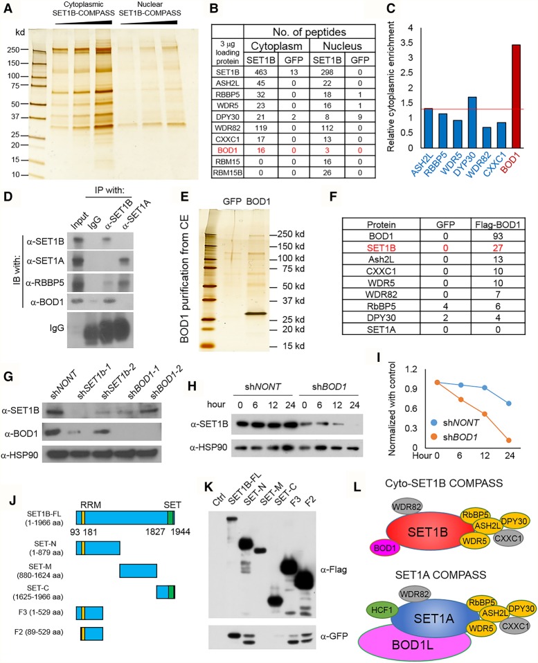
BOD1 is a cytoplasmic-specific subunit of SET1B/COMPASS. (A) MDA-MB-231 cells were stably infected with retroviruses expressing Flag-SET1B. Silver staining shows the Flag-tagged SET1B protein purified with M2 beads from either cytoplasmic extracts or nuclear extracts (equal volumes of eluted protein). (B) The mass spectrometry (MS) analysis with equal amounts of the interacting proteins of SET1B isolated from cytoplasmic and nuclear extracts are shown (equal amounts). (C) The results from B were normalized further with the spec count of the SET1B protein, and the relative cytoplasmic enrichment of the protein is shown. The cytoplasmic enrichment is presented as the ratio between cytoplasmic subunit/cytoplasmic SET1B and nuclear subunit/nuclear SET1B. (D) The endogen SET1A and SET1B proteins were immunoprecipitated, and the BOD1 protein in the immunoprecipitates was detected by Western blot. The common subunit of SET1A and SET1B COMPASS, RBBP5, was used as a control. n = 2. (E) Flag-BOD1 was purified from cytoplasmic extracts of MDA-MB-231-Flag-BOD1 cells and analyzed by silver staining. MS identified interacting proteins with Flag-BOD1. (F) Peptide numbers of subunits from SET1B COMPASS are shown. Cells stably expressing GFP were used as the negative control. (G) SET1B and BOD1 were knocked down with two distinct shRNAs in MDA-MB-231 cells, and the protein levels of SET1B and BOD1 were detected by Western blotting. HSP90 was used as the internal control. n = 3. (H) MDA-MB-231 cells were transfected with shBOD1 and selected by puromycin for 48 h. The cells were further treated with cycloheximide (CHX) for different times, and the protein level of SET1B was determined by Western blot. (I) The degradation ratios of SET1B in shNONT and shBOD1 cells were quantified by ImageJ. (J) A schematic diagram of SET1B full-length cDNA and truncated derivatives, each with a Flag tag fused to its N terminus, is shown. (K) Plasmids expressing the SET1B cDNA derivatives shown in J were transiently transfected into HEK293T cells together with GFP-BOD1 for 24 h. The Flag-tagged SET1B truncations were then purified, and the interacting GFP-BOD1 was detected by Western blotting with anti-GFP. n = 4. (L) Cartoon depictions of cyto-SET1B COMPASS and SET1A COMPASS. (Orange) WARD complex, which contains WDR5, ASH2L, RBBP5, and DPY30; (gray) SET1A/B COMPASS complex-specific subunits WDR82 and CXXC1; (pink) cyto-SET1B COMPASS-specific subunit BOD1.

