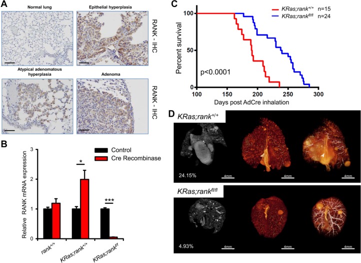
Figure 2.
Loss of RANK prolongs survival of KRasG12D -driven lung cancer. (A) Increased RANK expression during the progression of lung tumors. Analysis of RANK expression by IHC during lung tumor progression in KRasG12D mice. Representative images for RANK expression in atypical adenomatous hyperplasia (AAH), epithelial hyperplasia (EH), and adenoma of AdenoCre (AdCre)-infected mice are shown. Normal lung parenchyma is shown for a mouse not infected with AdCre (normal lung). AAH and EH were from mice 11 wk after AdCre infection; adenomas were from mice 14.5 wk after infection. Bars, 100 µm. (B) Quantitative PCR analysis of RANK mRNA levels in primary pneumocytes purified from KRas;rank+/+ and KRas;rankf/f mice; the experiment was performed with three technical replicates and repeated three times independently. Samples without AdCre infection in every genotype were set to 1, respectively. (*) P < 0.05; (***) P < 0.001, unpaired two-sided t-test. (C) Kaplan Meier survival curves for KRas;rank+/+ (n = 15; median survival 191 d) and KRas;rankfl/fl (n = 24; median survival 239 d) littermate mice injected intranasally with AdCre (2.5 × 10⁷ plaque-forming units). P < 0.0001 (log rank test) between KRas;ranklfl/fl and littermate controls. (D) Representative microCT images of lung tumors of KRas;rank+/+ and KRas;rankfl/fl littermate mice assayed 25 wk after AdCre inhalation. Three different images of the same lung are shown, and the percentages of tumor to lung volumes are indicated. See also Supplemental Figure S3A.

