Figure 4.
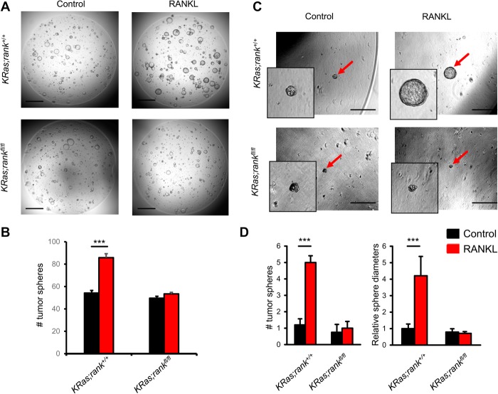
RANK drives lung cancer stem-like cell expansion. (A) Representative images of tumor spheroids derived from purified KRas;rank+/+ and KRas;rankfl/fl primary lung tumor cells. Images were acquired 4 d after 1 µg/mL RANKL treatment. Five-thousand primary tumor cells were seeded per plate. The experiment was designed with six replicates for each condition and repeated with three different mice for each group, respectively. Bars, 1 mm. (B) Quantitative analysis (mean ± SEM) of tumor spheroid numbers described in A. (***) P < 0.001, unpaired two-sided t-test. (C) Representative images of tumor spheroids (red arrows) derived from purified KRas;rank+/+ and KRas;rankfl/fl primary lung tumor cells. Images were acquired 7 d after 1 µg/mL RANKL treatment. Five-hundred primary tumor cells were seeded per plate. The experiment was designed with six replicates for each condition and repeated with three different mice for each group, respectively. Bars, 500 μm. (D) Quantitative analysis (mean ± SEM) of tumor spheroid numbers (left panel) and relative tumor spheroid diameters (right panel). The average diameters of tumor spheroids in KRas;rank+/+ without RANKL treatment was arbitrarily set as 1 to normalize all other groups. (***) P < 0.001, unpaired two-sided t-test.

