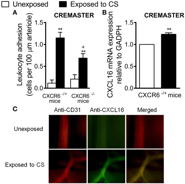
Figure 7.

Effect of cigarette smoke (CS) exposure in CXCR6-expressing and CXCRC6 knockout mice. Heterozygous (CXCR6−/+) and homozygous (CXCR6−/–) mice were exposed or not to CS for 3 days and responses were examined 16 h later. (A) Leukocyte–arteriolar endothelium interactions was measured by intravital microscopy. Results are expressed as mean ± SEM (n = 5–8 animals per group). *P < 0.05 or **P < 0.01 relative to non-exposed animals; +P < 0.05 relative to CXCR6−/+ mice. (B) Relative quantification of CXCL16 and β-actin mRNA was determined by RT-PCR. Columns show fold increase in expression of CXCL16 mRNA relative to control GAPDH values (n = 5 independent experiments). Values are represented as mean ± SEM of the 2−ΔΔCt values. **P < 0.01 relative to non-exposed animals. (C) Cremaster muscle was fixed for CXCL16 and endothelium (CD31) staining. CXCL16 expression is shown in green (stained with an Alexa Fluor 488-conjugated donkey anti-rabbit secondary antibody) and vessel endothelium (red) was stained with a PE-conjugated anti-mouse CD31 monoclonal antibody. Overlapping expression of CXCL16 and CD31 is shown in yellow. Results are representative of five to six animals per group.
