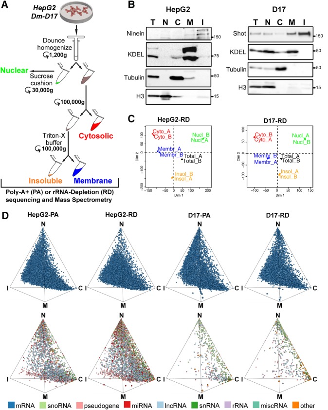
FIGURE 1.
Cell fractionation combined with RNA sequencing (CeFra-seq) of human and Drosophila epithelial cell models. (A) Schematic diagram of the fractionation procedure based on Dounce homogenization, centrifugation, and detergent extraction steps to obtain nuclear, cytosolic, membrane, and insoluble fractions. (B) Western blots of protein sample controls show fraction efficiency. The accumulation of the indicated protein markers was assessed in human HepG2 and Drosophila D17 cells. (C) Principal component analysis of RNA-seq replicates for HepG2 and D17 cells. (D) Simplex graph of the relative localization of mRNAs (top row) or noncoding RNAs (bottom row) across subcellular fractions, either assessed from poly(A)-enriched (PA) or rRNA-depleted (RD) sequencing data sets. (T) Total, (C) cytosolic, (M) membrane, (I) insoluble, (N) nuclear.

