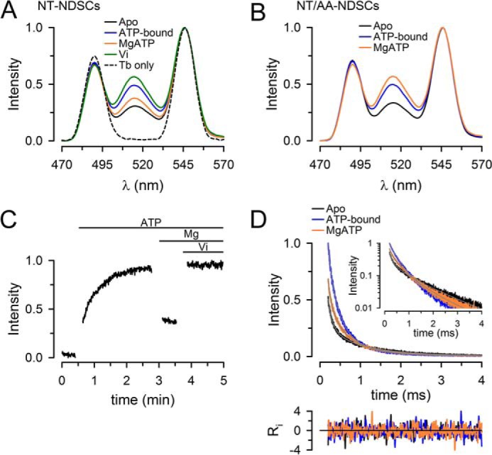
Figure 3.

Conformational changes during the ATP hydrolysis cycle of NT Pgp in NDSCs at 37 °C. A, emission spectra from NDSCs containing NT Pgp labeled with donor only (Tb3+ chelate) or donor and acceptor (Tb3+ chelate and Bodipy FL). Traces were normalized to the Tb3+ emission at 546 nm. B, emission spectra of the catalytically-inactive mutant NT/AA Pgp in NDSCs. C, time course of the changes in Bodipy FL-sensitized emission, recorded at 520 nm, in response to sequential additions of NaATP, MgSO4, and Vi. The gaps during the recording correspond to time periods where additions and manual mixing were performed. The intensity was normalized to the maximum obtained after addition of Vi. D, Bodipy FL sensitized emission decays, recorded at 520 nm, from NT Pgp in NDSCs. The inset displays the same curves in a semi-log scale. Ri represents the weighted residuals of the multiexponential fits (gray lines in the main graph and inset). Decays were normalized to the emission of the ATP-bound protein at 200 μs. All the traces in the figures are representative of at least seven independent experiments and were obtained at 37 °C. Emission was recorded after a 200-μs delay from the 337-nm excitation pulse. Apo, nucleotide- and drug-free buffer with 1 mm EDTA; ATP-bound, + 5 mm NaATP; MgATP, +10 mm MgSO4.
