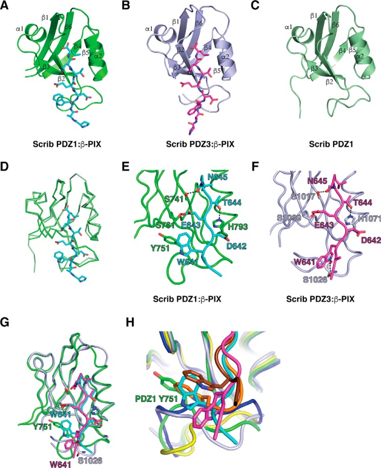
Figure 5.
Crystal structures of PDZ1 and PDZ3 each bound to a β-PIX peptide. The β-PIX peptide engages individual PDZ domains via a shallow groove located between the β2 and α2. A, PDZ1 (green) is shown as a schematic with β-PIX peptide (cyan) represented as sticks. B, PDZ3 (light purple) is shown as a schematic with β-PIX peptide (magenta) represented as sticks. C, ligand-free PDZ1 is shown as a schematic (forest green). D, overlay of ribbon traces of PDZ1 with (green) and without β-PIX peptide (forest green). E and F, PDZ1 and -3 domains bound to β-PIX peptides are shown as ribbons and sticks, respectively. Side chains of the residues involved in interactions (shown as dashed black lines) are displayed as sticks and are labeled. G, superimposition of PDZ1 and PDZ3 bound to β-PIX peptide. H, superimposition of Scribble PDZ1 (green and cyan) and PDZ3 (light purple and magenta) complexes with β-PIX peptide and Erbin (dark blue and chocolate, PDB code 1N7T) and ZO-1 (yellow and orange, PDB code 2H2C) complexes with synthetic peptides.

