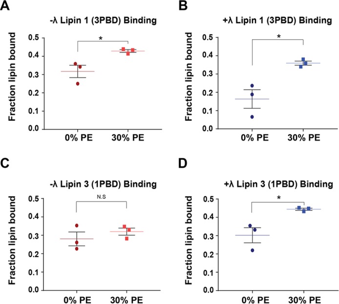
Figure 6.

Binding of Venus-lipin PBD exchange mutants with PA containing liposomes. Phosphorylated (−λ) Venus-tagged lipin 1 (3PBD) (A), dephosphorylated (+λ) Venus-tagged lipin 1 (3PBD) (B), −λ Venus-tagged lipin 3 (1PBD) (C), and +λ Venus-tagged lipin 3 (1PBD) (D) were subjected to liposome floatation assays using PC/PA and PC/PE/PA (30 mol % PE) liposomes, containing 20 mol % PA and 0.1 mol % PC-pyrene. The final concentration of PA in all assays was 2 mm. Shown is a scatter plot with a mean of triplicate determinations from one representative experiment ± S.E. (error bars). Student's t test was used to analyze statistical analysis. *, p < 0.05.
