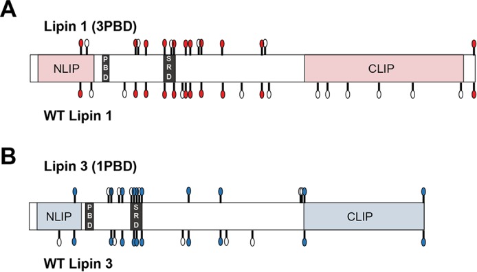
Figure 8.

Shared phosphorylated sites among WT lipins 1 and 3 and the PBD mutants. Shown is a schematic of the phosphorylation sites found in the four indicated proteins. The white circles indicate the position of the phosphorylation sites unique to the given protein. The colored circles indicate the position of the phosphorylation sites shared between WT lipin 1 and lipin 1 (3PBD) (A) and WT lipin 3 and lipin 3 (1PBD) (B). CLIP and NLIP, C-terminal and N-terminal domain, respectively.
