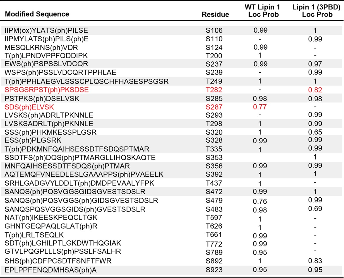
Table 4.
WT lipin 1 and lipin 1 (3PBD) phosphorylation analysis by LC-MS/MS
Rows highlighted in grey indicate phosphorylated residues with localization probabilities of ≥ 0.95 in both proteins. Localization probabilities are left blank if no peptide was detected or if the localization probability was < 0.5. With the exception of the sites in red, sites are only reported if they have localization probabilities ≥ 0.95 in at least one of WT lipin 1 or lipin 1 (3PBD). Sites in red are those believed to be important in lipin activity and regulation but were not detected with a localization probability of ≥ 0.95 in either protein. However, we have > 99% confidence that there is a phosphorylation site in these peptide regions. Peptide sequences come from the WT protein unless the WT localization probability is < 0.95, in which case the peptide corresponding to the highest site localization probability is shown. Only sites with high peptide identity confidence (PEP ≤ 0.05) are listed.

