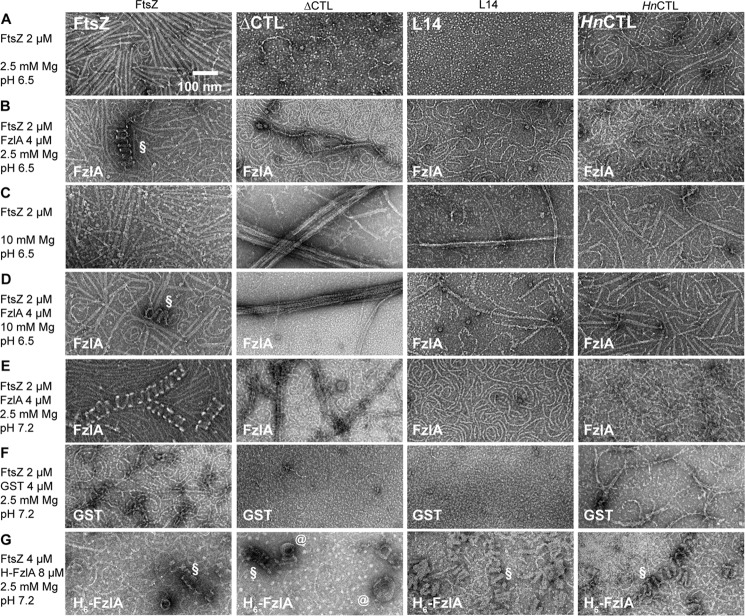
Figure 12.
FzlA-induced bundling of FtsZ protofilaments into helices is CTL-dependent. A–E, electron micrographs of polymers formed by 2 μm FtsZ CTL variants in the presence or absence of 2 μm FzlA at different pH (6.5 versus 7.2) with 2.5 or 10 mm MgCl2, 50 mm KCl, and 2 mm GTP spotted on grids 15 min after addition of nucleotide and stained with uranyl formate. F, electron micrographs of polymers formed by 2 μm FtsZ CTL variants in the presence of 2 μm glutathione S-transferase (GST) at pH 7.2 with 2.5 mm MgCl2, 50 mm KCl, and 2 mm GTP spotted on grids 15 min after addition of nucleotide and stained with uranyl formate. G, electron micrographs of polymers formed by 4 μm FtsZ CTL variants in the presence of 8 μm His6-FzlA at pH 7.2 with 2.5 mm MgCl2, 50 mm KCl and 2 mm GTP spotted on grids 15 min after addition of nucleotide and stained with uranyl formate. Scale bar, 100 nm. §, helical bundles of FtsZ (or CTL variant) with FzlA; @, spiral structures seen for ΔCTL with His6-FzlA.

