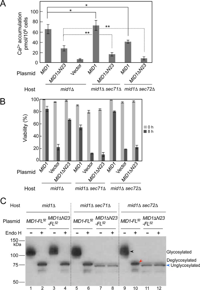
Figure 6.
Mid1ΔN23 is translocated to the ER post-translationally. Shown are Ca2+ accumulation (A) and the viability (B) of the mid1Δ, mid1Δ sec71Δ, and mid1Δ sec72Δ mutants transformed with the plasmid YCpS-MID1 or YCpS-MID1ΔN23. Cells of the transformants were treated with α-factor and subjected to the measurement of Ca2+ accumulation and cell viability as described in the legend to Fig. 3. C, identification of N-glycosylation of the MID1ΔN23-FL52 protein with the Endo H treatment. The mid1Δ, mid1Δ sec71Δ, and mid1Δ sec72Δ mutants were transformed with the plasmid YCpS-MID1-FL52 or YCpS-MID1ΔN23-FL52. Crude extracts prepared from the transformants were subjected to the Endo H treatment, followed by SDS-PAGE and Western blotting, as described in the legend to Fig. 4, except that an anti-FLAG antibody was used to detect the proteins to be identified. The amount of whole proteins in the crude extracts loaded to lanes 7, 8, 11, and 12 was 5 times as high as that loaded to other lanes. Black arrowhead, N-glycosylated protein; red arrowhead, deglycosylated protein; blue arrowhead, unglycosylated protein. For simplicity, arrowheads are shown only in lanes 9–12. Error bars, S.D.

