
Figure 8.
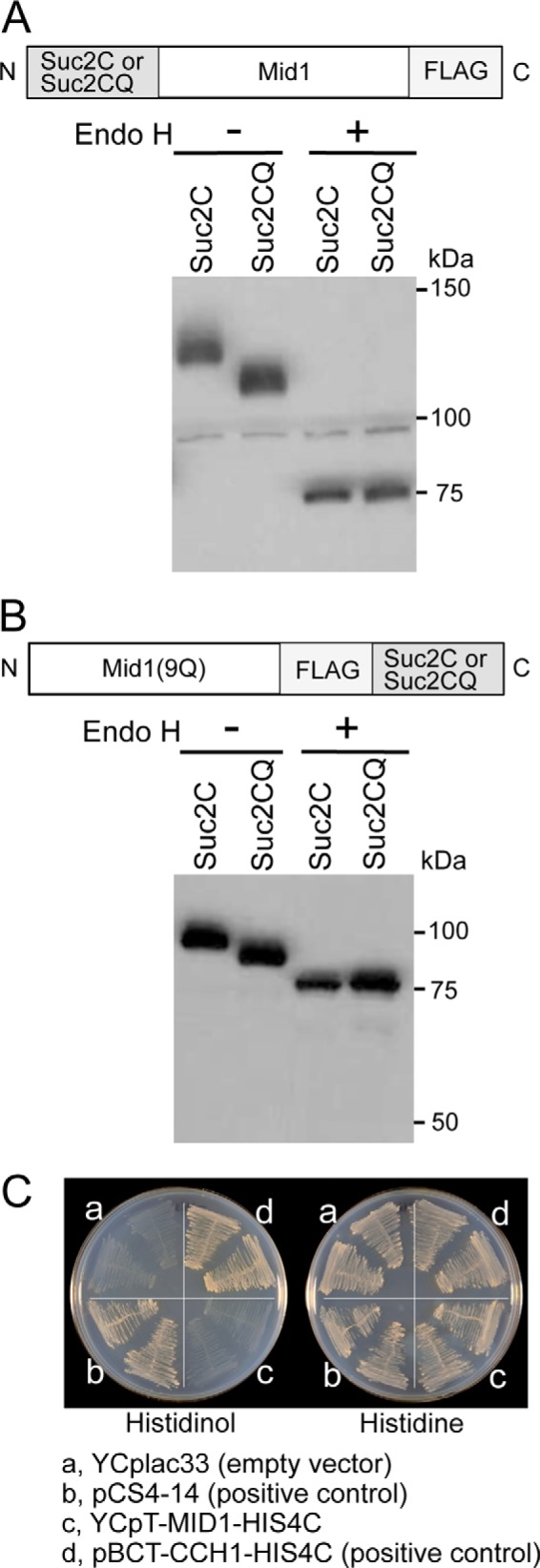
N and C termini of Mid1 are present outside the cell and in the ER lumen. A, subcellular localization of the N terminus of Mid1. The Mid1 protein tagged N-terminally with the N-glycosylation reporter Suc2C or its negative control Suc2CQ was treated with Endo H or mock-treated and then subjected to Western blotting with an anti-FALG antibody. B, subcellular localization of the C terminus of Mid1. The Mid1(9Q) protein tagged C-terminally with Suc2C or Suc2CQ was subjected to the same treatment and analysis as described above. C, histidinol plate assays. The yeast strain FC2a transformed with each of the following four plasmids was streaked onto a histidinol plate (left) and histidine plate (right), incubated at 30 °C for 3 days, and then photographed. a, YCplac33 (empty vector; negative control); b, pCS4–14 (positive control); c, YCpT-MID1-HIS4C; d, pBCT-CCH1-HIS4C (positive control).