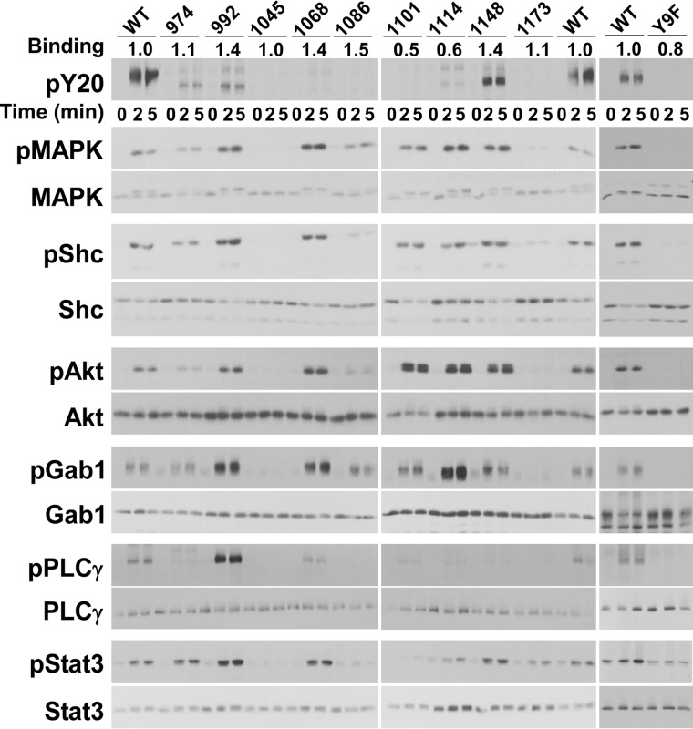
Figure 3.
Signaling by the single-Tyr receptors. Cells stably expressing wild-type or one of the single-Tyr EGF receptors were treated with 10 nm EGF for 1, 2, or 5 min at 37 °C as indicated. RIPA lysates were prepared and separated by SDS-PAGE. For the anti-phosphotyrosine blots, the protein load for the wild-type receptor was one-third that of the single-Tyr receptors. For all other blots, equal protein was loaded for the wild-type and the single-Tyr receptors. The values at the top of the Western blots in the line labeled Binding represent the binding of 125I-cetuximab to each cell line in that experiment, normalized to that observed for cells expressing the wild-type EGF receptor.

